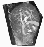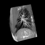

In contrast to CT, MRI provides excellent soft tissue contrast and volunteers and patients are not exposed to ionising radiation.
Sequences of 3D volumes (4D data sets) were reconstructed from dynamic sagittal 2D images acquired during free breathing. Other gating methods assume regular respiratory motion and reduce the respiratory organ deformation to one parameter such as amplitude or phase. This neglects all residual variability and is a too coarse approximation in some cases, leading to artefacts in the reconstructed images.
The proposed approach derives a multi-dimensional gating measure from dedicated so-called navigator frames in order to determine the state of the liver retrospectively and find corresponding 2D slices that can be combined to 3D volumes. The method does not assume a constant breathing depth or even strict periodicity and does not depend on an external gating signal. The technique is applicable to any organ that undergoes respiratory motion such as lung, liver, pancreas or kidneys and can be implemented on a standard MR scanner without additional equipment.
 |
 |
| animate | animate |
| Example sequences showing typical breathing cycles reconstructed from free breathing sagittal slices. | |